All About Ultrasounds
You’ve probably heard that ultrasounds are used during pregnancy, but if you’ve never had one before, there can be lots of questions! This article is all about ultrasounds and answering those questions.
What is an Ultrasound?
An ultrasound scan is a way of using sound waves to capture images of the inside of a body. While very commonly used in pregnancies to see images of the baby, ultrasounds are also used to view organs and movement within them. Ultrasounds are also called sonograms because the process involves transmitting sound waves at a high frequency to create an image. It is noninvasive and provides your doctor with lots of valuable information.
Why are Ultrasounds Used in Pregnancy?
There are a number of reasons to have an ultrasound at various stages of pregnancy. One can be performed as early as 6-10 weeks to both confirm the pregnancy and determine an estimated due date. However, this is typically performed between 12 and 16 weeks.
The physician can also track the progress of your growing baby. He or she will be able to see any fetal abnormalities at 18-22 weeks. The physician will also listen to your baby’s heartbeat to make sure that it is normal. This is also the time that the baby’s sex can typically be determined, if you want to know. However, there are situations in which the baby is not in the right position to identify the sex.
Closer to the time of delivery, the doctor can determine if the baby is in the correct position for birth and look out for any abnormalities that could potentially complicate delivery.
What does the Process Look Like?
There are two types of ultrasounds: the pelvic and the transvaginal.
A pelvic ultrasound is noninvasive and not painful. The doctor will spread a clear, lubricating gel on the skin to help improve transmission of the sound waves. The gel will also help the transducer to travel smoothly over your skin. It will release high-frequency sound waves through your body, which will echo when they hit a dense object. These echoes are transferred back to a computer which can translate them into a video monitor so that you can see your baby!
The transvaginal ultrasound is often used during the first trimester because it provides better images. In this type of ultrasound, the transducer is in the shape of a small wand. It will be covered with a latex sheath and inserted into the vagina. The transducer will be moved around slightly to form a clear picture of the baby.
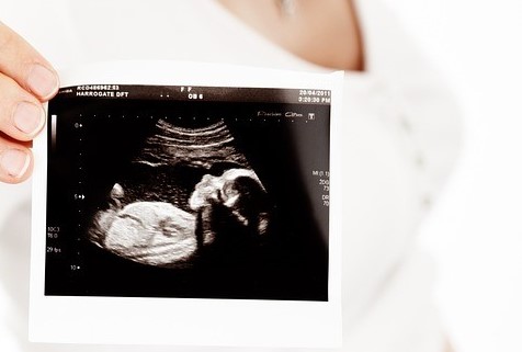
The basic ultrasound process generally takes about 15-20 minutes. Afterwards, you and your doctor can review the images, and they may give you a printout to take home as a keepsake. They may call you later to discuss any findings or schedule a follow-up appointment.
The bottom line is that an ultrasound is a very useful test to learn more about your baby and periodically check that everything is progressing as expected. Cheyenne OBGYN is proud to be accredited by the American Institute of Ultrasound in Medicine and is currently the only accredited ultrasound department for obstetrical and gynecologic ultrasound in the state of Wyoming. We’d be honored to perform your ultrasounds during your pregnancy. Please call or text us at 307-634-5216 if you have any other questions regarding ultrasounds!
Leave a Reply